
Radiology
Radio logy
Radiology for iMed Billing
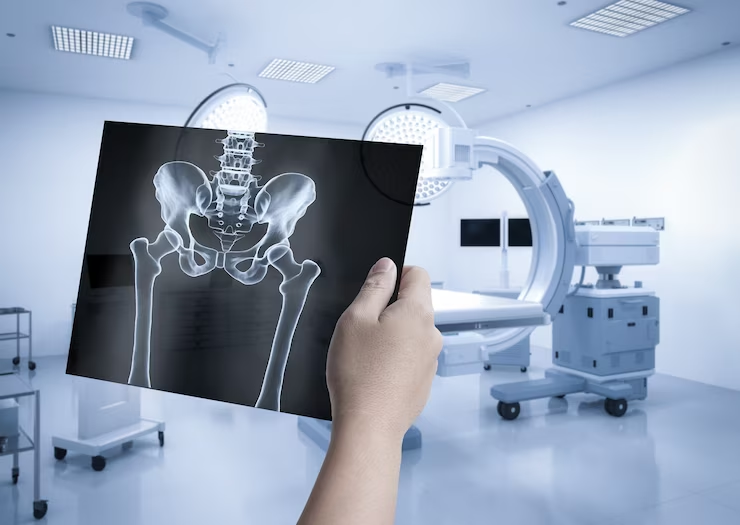
Radiology is a medical specialty that dealt with imaging technologies to detect/diagnose and treat diseases in humans and animals. It originated with X-rays, which use ionizing radiation to capture images of bones and tissues. Over time, radiology has expanded to encompass a wide array of imaging modalities, both those that use ionizing radiation and those that do not.
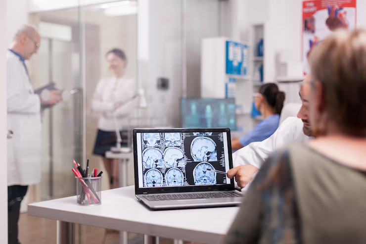
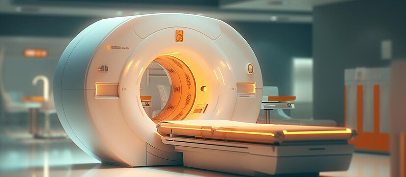
X-rays remain fundamental, primarily for detecting bone fractures, infections, and tumors. Computed Tomography (CT) scans provide detailed body cross-sectional pictures created by fusing many X-ray pictures, enhancing the visibility of bones, blood vessels, and soft tissues. Magnetic Resonance Imaging (MRI), which uses strong magnets and radio waves, generates high-resolution images of organs and tissues without ionizing radiation, making it ideal for brain, spine, and joint imaging.
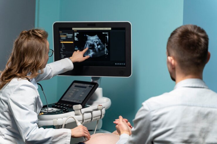
Ultrasonography employs high-frequency sound waves to create real-time images of internal organs, blood flow, and developing fetuses, offering a radiation-free alternative. Fluoroscopy allows continuous X-ray imaging to observe internal structures in real-time and is commonly used in procedures like cardiac catheterization and gastrointestinal studies

Radio Logy
Nuclear Medicine small doses of radioactive elements, or radiotracers, are used in nuclear medicine to diagnose and cure illnesses. Methods such as Single-Photon-Emission-Computed-Tomography (SPECT) and Positron-Emission-Tomography (PET) offer fine-grained imaging of the body's physiological and metabolic processes. .
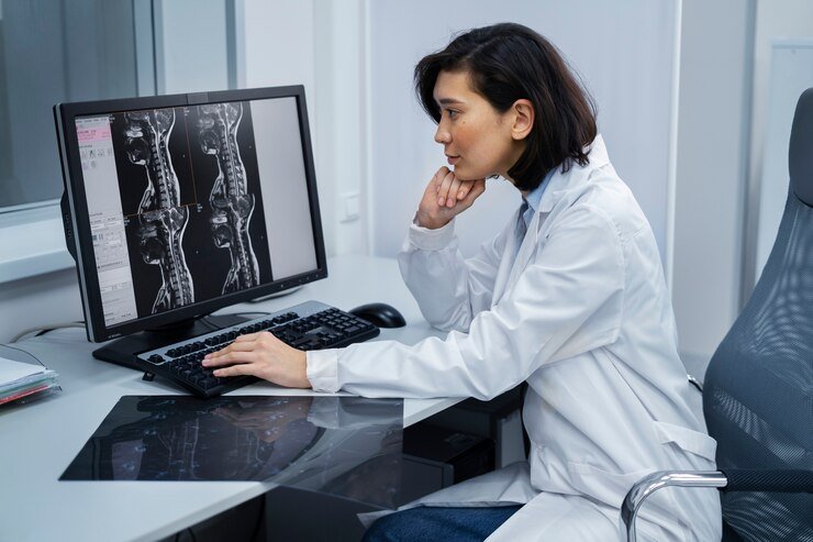
Radiologists, medical doctors specializing in interpreting these images, play a crucial role in diagnosing conditions and guiding treatment plans. Radiologic technologists operate imaging equipment and ensure high-quality image capture

Radio Logy
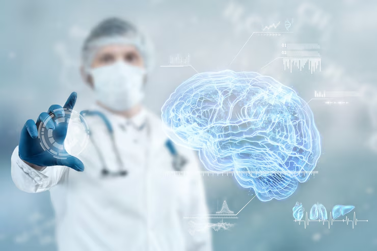
Digital imaging has advanced significantly, allowing for better image quality and more accessible storage and sharing. Additionally, Artificial Intelligence (AI) is increasingly used to assist in image analysis, improving diagnostic accuracy and efficiency.
Radiology's integration of various imaging techniques continues to enhance the ability to diagnose and treat multiple medical conditions effectively and safely, ensuring comprehensive patient care
Key Imaging Modalities in Radiology
Radiology's integration of various imaging techniques continues to enhance the ability to diagnose and treat multiple medical conditions effectively and safely, ensuring comprehensive patient care

Detailed Components
X-rays
The first and most well-known form of medical imaging. They are primarily used to view bones and detect fractures, infections, or tumors. Provides quick, inexpensive, and efficient diagnostic images
Computed Tomography
Combines multiple X-ray pictures taken from different angles to produce cross-sectional body views. Provides detailed images of bones, blood vessels, and soft tissues. Essential for diagnosing complex conditions like cancers, cardiovascular diseases, and traumatic injuries.
Magnetic Resonance Imaging
Develops clear pictures of organs and tissues using radio waves & strong magnets tissues. Does not use ionizing radiation, making it safer for frequent use. Ideal for brain, spine, joint, and soft tissue imaging
Ultrasonography
Utilizes high-frequency-sound-waves to produce real-time images. It is commonly used to examine internal organs, monitor blood flow, and check fetal development during pregnancy.
Detail Components
Radiology's integration of various imaging techniques continues to enhance the ability to diagnose and treat multiple medical conditions effectively and safely, ensuring comprehensive patient car
Fluoroscopy
Provides continuous X-ray imaging, allowing real-time observation of internal organs. They are often used in procedures like barium X-rays and cardiac catheterization. Helpful in guiding interventional procedures and diagnosing functional abnormalities.
Nuclear Medicine
Involves using small amounts of radioactive materials (radiotracers) to diagnose and treat diseases. Techniques such as Positron-Emission-Tomography (PET) and single-photon emission computed tomography (SPECT) provide metabolic and functional information.

Functions and Applications
-
Diagnosis
Radiology is crucial in detecting and diagnosing various conditions, from fractures and infections to cancers and cardiovascular diseases. Provides critical information that guides patient management and treatment decisions. Treatment: Interventional radiology is a subspecialty where imaging techniques guide minimally invasive surgical procedures. Procedures such as angioplasty, stent placement, and biopsy are performed precisely using imaging guidance.
-
Monitoring and Follow-up
Radiologists track the progress of diseases and the effectiveness of treatments through periodic imaging studies. Ensures accurate and timely medical care and adjustments to treatment plans as needed
-
Research
Advances in imaging technologies and techniques continually expand the capabilities of radiology. Contributes to medical research and the development of new diagnostic and therapeutic methods

Radiology Professionals
Radiology's integration of various imaging techniques continues to enhance the ability to diagnose and treat multiple medical conditions effectively and safely, ensuring comprehensive patient care

Detailed Components
Radiologists
Medical doctors specialize in interpreting medical images, diagnosing conditions, and recommending appropriate treatments. Often dealt with other specialists to provide comprehensive care
Radiologic Technologists
Skilled professionals who operate imaging equipment, prepare patients for procedures and ensure high-quality images for accurate diagnosis. Play a crucial role in the imaging process and patient care. Technological Advancements
Digital Imaging
It enhances image quality and allows for easy storage, retrieval, and sharing of medical images. Facilitates better communication among healthcare providers.
Artificial Intelligence
AI algorithms assist in image analysis, improving accuracy, efficiency, and early detection of abnormalities. Enhances diagnostic capabilities and supports clinical decision-making.
Detail Components
Hybrid Imaging
Combines multiple imaging modalities (e.g., PET-CT) to provide more comprehensive diagnostic information. Integrates functional and anatomical imaging for better diagnosis and treatment planning
Radiation Safety
Radiologists and technologists prioritize minimizing radiation exposure to patients. Techniques like dose optimization and protective shielding are used to ensure patient safety.
Ethical Practice
Ensuring patient consent, privacy, and the ethical use of imaging technologies is paramount in radiological practice. Upholds professional standards and fosters trust between patients and healthcare providers


Benefits of Radiology
-
Accurate Diagnosis
Radiology provides detailed and precise images of the body's internal structures, aiding in accurately diagnosing various medical conditions, from fractures to complex diseases like cancer and cardiovascular issues.
-
Early Detection
Imaging techniques like that: mammography, CT scans, and MRIs allow for the early detection of diseases, which is crucial for conditions like cancer, where early intervention can significantly improve outcomes
-
Noninvasive
Many radiological procedures, such as ultrasound and MRI, are noninvasive, meaning they do not need surgery or the insertion of instruments into the body. This reduces patient discomfort and recovery time
-
Real-Time Imaging
Modalities like fluoroscopy provide real-time imaging, invaluable during surgical procedures and in diagnosing dynamic processes within the body, such as blood flow and organ function
-
Comprehensive Evaluation
Radiology allows for a comprehensive evaluation of the body's internal organs and systems, often providing a complete picture of a patient's health compared to traditional diagnostic methods


Detailed Components
Treatment Planning
Detailed imaging is essential for planning surgical procedures, guiding minimally invasive surgeries, and ensuring precision in radiation therapy for cancer treatment, thereby improving treatment outcomes
Monitoring Progress
Radiologists can monitor the progress of diseases and the effectiveness of treatments over time, helping to adjust therapeutic approaches as needed for better patient outcomes.
Pain Management
Interventional radiology techniques can be used to manage chronic pain, such as through image-guided injections and nerve blocks, providing relief for patients with conditions like arthritis or back pain
Increased Safety
Modern radiological techniques are designed to minimize exposure to ionizing radiation, making diagnostic imaging safer for patients. Additionally, non-ionizing modalities like MRI and ultrasound do not expose patients to radiation
-
Accessibility
Radiological services are widely available in hospitals, clinics, and specialized imaging centers, making it easier for patients to access necessary diagnostic and therapeutic procedures
-
Enhanced Patient Care
Radiology supports a multidisciplinary approach to patient care, with radiologists working closely with other healthcare providers to deliver comprehensive and coordinated treatment plans.
-
Cost-Effective
By providing early and accurate diagnosis, radiology can help reduce healthcare costs by avoiding unnecessary surgeries, minimizing hospital stays, and preventing the progression of diseases through timely intervention.
-
Patient Education
Visual aids from radiological images help healthcare providers explain medical conditions to patients, improving understanding and enabling informed decision-making about their care
-
Technological Advancements
Continuous advancements in imaging technology, such as digital imaging, AI integration, and hybrid imaging techniques, enhance diagnostic accuracy, efficiency, and patient outcomes.


Preventive Health
Routine imaging screenings, such as mammograms and DEXA scans for bone density, play a vital role in preventive health. They identify potential issues before they develop into severe conditions.
Improved Quality of Life:
By enabling early and accurate diagnosis, effective treatment planning, and ongoing monitoring, radiology contributes significantly to enhancing the overall excellence of life for patients
CPT Codes for Radiology
Diagnostic Radiology (X-rays)
Computed Tomography (CT)
CT scan, head or brain; without contrast
CT scan, head or brain, with contrast material(s)
CT scan, thorax; without contrast
CT scan, thorax; with contrast material(s)
CT scan, cervical spine; without contrast
CT scan, lumbar spine; without contrast
CT scan, abdomen; without contrast
CT scan, abdomen; with contrast material(s)
CT scan, abdomen and pelvis; without contrast
Magnetic Resonance Imaging
MRI, brain; without contrast material(s)
MRI, brain; without and with contrast material(s)
MRI, cervical spine; without contrast
MRI, lumbar spine; without contrast
MRI, upper extremity joint (e.g., shoulder, elbow, wrist); without contrast
MRI, lower extremity joint (e.g., hip, knee, ankle); without contrast
MRI, abdomen; without contrast
MRI, abdomen; without and with contrast material(s)
Ultrasonography (Ultrasound)
Ultrasound, breast; unilateral, complete
Ultrasound, abdomen; complete
Ultrasound, obstetric; first trimester, less than 14 weeks gestation, transabdominal approach; single or first gestation
Ultrasound, obstetric; greater than or equal to 14 weeks 0 days gestation, transabdominal approach; single or first gestation
Ultrasound, pelvic (nonobstetric); complete
Ultrasonic guidance for needle placement (e.g., biopsy, aspiration, injection, localization device), imaging supervision and interpretation
Fluoroscopy (separate procedure), up to 1 hour
Fluoroscopic guidance for vascular access and catheter manipulation, any necessary contrast injections through the access site or catheter with associated venography radiologic supervision and interpretation, and radiographic documentation of the final catheter position are all included in the placement, replacement, or removal of central venous access devices.
Fluoroscopic guidance for needle placement (e.g., biopsy, aspiration, injection, localization device), imaging supervision and interpretation
Nuclear Medicine
Thyroid uptake; single or multiple determinations
Gastric emptying study, with small bowel transit
Myocardial perfusion imaging, tomographic (SPECT); multiple studies, at rest and stress
Concurrently obtained CT and positron emission tomography (PET) for attenuation correction and anatomical localization imaging (skull base to mid-thigh)
Revascularization, endovascular, open or percutaneous, iliac artery, unilateral; initial vessel
Interventional Radiology
Revascularization, endovascular, open or percutaneous, femoral/popliteal artery, unilateral; with atherectomy, includes angioplasty within the same vessel, when performed
Endovascular stent placement, open or percutaneous, iliac artery, each vessel; radiological supervision and interpretation
Screening mammography, bilateral (2-view study of each breast), including computer-aided detection (CAD) when performed
Diagnostic mammography, including CAD when performed; unilateral
Diagnostic mammography, including CAD when performed; bilateral

At iMedbillingPro, we are aware of the challenges and suffering that healthcare providers encounter when delivering first-rate patient care.
Copyright © 2024 iMed Billing Pro All Rights Reserved.